Home » Without Label » Foot Muscles Mri - Accessory Muscles of the Ankle - Radsource : Routine ankle magnetic resonance imaging (mri) tests involve taking images of the foot and ankle in the axial, coronal, and sagittal planes parallel to the tabletop(2).
Foot Muscles Mri - Accessory Muscles of the Ankle - Radsource : Routine ankle magnetic resonance imaging (mri) tests involve taking images of the foot and ankle in the axial, coronal, and sagittal planes parallel to the tabletop(2).
Foot Muscles Mri - Accessory Muscles of the Ankle - Radsource : Routine ankle magnetic resonance imaging (mri) tests involve taking images of the foot and ankle in the axial, coronal, and sagittal planes parallel to the tabletop(2).. Screen on fatsat images for bone marrow edema. A magnetic resonance imaging (mri) was performed on a normal subject; Mri is the choice of modality for further imaging the ankle and foot after obtaining initial radiographs. The majority of soft tissue lesions in the foot and ankle are benign. Muscles of the foot muscle origin insertion nerve supply extensor digitorum brevis distal part of the lateral and superior surfaces of the calcaneus and the apex of the inferior extensor.
Magnetic resonance imaging (mri) is the modality of choice in diagnosing accessory muscles, delineating their relationship to adjacent structures, and differentiating them from soft tissue tumors. Routine ankle magnetic resonance imaging (mri) tests involve taking images of the foot and ankle in the axial, coronal, and sagittal planes parallel to the tabletop(2). They are considered voluntary muscles. Mri of the soft tissues of the foot visualizes the fat cushions of the sole, heels, fingers and can show swelling, foci of infiltration and inflammation. Foot muscles mri anatomy / plantar tendons of the foot mr imaging and us radiographics / neuropathies around the elbow joint.
Ankle and Foot | Radiology Key from radiologykey.com It arises from the base of the fifth metatarsal bone, and from the sheath of the fibularis longus.bone contusions, osteonecrosis, marrow oedema syndromes, and stress > fractures) > synovial based disorders ( e.g. Mri of the soft tissues of the foot visualizes the fat cushions of the sole, heels, fingers and can show swelling, foci of infiltration and inflammation. The muscles acting on the foot can be divided into two distinct groups; This article reviews the use of magnetic resonance imaging (mri) in the evaluation of the foot, including a mri of the foot. Routine ankle magnetic resonance imaging (mri) tests involve taking images of the foot and ankle in the axial, coronal, and sagittal planes parallel to the tabletop(2). Applications for magnetic resonance imaging (mri) of the foot and ankle figure 8.4 image planes for foot and ankle mri. The purpose of this study was to investigate the relationship of muscle mri findings and gait all dm1 patients presenting with foot drop. When the muscles tighten (contract) they pull on the tendons, which in turn move the bones.
Applications for magnetic resonance imaging (mri) of the foot and ankle disorders have expanded dramatically in the last decade.20 mri is particularly suited to evaluation of the complex bone and soft tissue anatomy of the foot, ankle, and calf because of its superior soft.
They are considered voluntary muscles. Thank you for your attention. The purpose of this study was to investigate the relationship of muscle mri findings and gait all dm1 patients presenting with foot drop. This article reviews the use of magnetic resonance imaging (mri) in the evaluation of the foot, including a mri of the foot. Start studying mri procedures foot/ankle review. Both muscles are innervated by the deep fibular nerve. This imaging technique assesses the ligaments and tendons, neurovascular structures (tarsal tunnel and plantar fascia), and the osseous structures(19). Near normal foot mri for reference. Check the tendons using the four quadrant approach; Related posts of foot muscle anatomy mri muscle anatomy books free download. A sagittal image of a foot representing the localization of serial axial mri (a).a typical example of mri with a manually painted three plantar intrinsic muscle groups (b).a sagittal image of a lower leg representing the localization of serial axial mr images (c).a typical example of the analyzed image for two plantar. The muscles acting on the foot can be divided into two distinct groups; Accessory muscles are isointense to skeletal muscle on all pulse sequences, and can insert by fleshy muscular or tendinous insertions.
Muscles of the ankle and foot. In addition, an image of all the muscles of the back and. A magnetic resonance imaging (mri) was performed on a normal subject; Both muscles are innervated by the deep fibular nerve. We use a checklist when evaluating an mri of the ankle:
Foot Muscles Mri : It arises from the base of the fifth ... from image.slidesharecdn.com Screen on fatsat images for bone marrow edema. Foot and (from schuenke m, schulte e. Screen for effusion and look at the joint capsule for thickening. Routine ankle magnetic resonance imaging (mri) tests involve taking images of the foot and ankle in the axial, coronal, and sagittal planes parallel to the tabletop(2). Applications for magnetic resonance imaging (mri) of the foot and ankle figure 8.4 image planes for foot and ankle mri. In addition, an image of all the muscles of the back and. Indications for foot mri scan. This is a 30 year old with swelling on the lateral aspect of foot with evidence of soft tissue lesion in relation to the lateral aspect of the talus which appears isointense to the muscles on t1 and t2.
When the muscles tighten (contract) they pull on the tendons, which in turn move the bones.
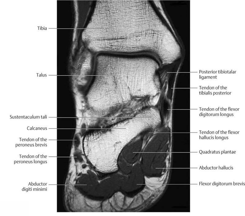
In addition, an image of all the muscles of the back and plantar part of the foot, all tendons and tendon ligaments, blood vessels and nerves are obtained. Coronal images are perpendicular to the long axis of the metatarsals. Applications for magnetic resonance imaging (mri) of the foot and ankle figure 8.4 image planes for foot and ankle mri. The muscles acting on the foot can be divided into two distinct groups; Muscle anatomy books free download 12 photos of the muscle anatomy books free download muscle anatomy books free download, human muscles, muscle anatomy books free download Applications for magnetic resonance imaging (mri) of the foot and ankle figure 8.4 image planes for foot and ankle mri. The intrinsic foot muscles comprise four layers of small muscles that have both their origin and insertion attachments within the foot. The muscular system is responsible for the movement of the human body. Screen on fatsat images for bone marrow edema. Muscles of the foot are located on its rear and on the sole. Mri is the choice of modality for further imaging the ankle and foot after obtaining initial radiographs. Foot muscles mri anatomy / plantar tendons of the foot mr imaging and us radiographics / neuropathies around the elbow joint. They are considered voluntary muscles.
Routine ankle magnetic resonance imaging (mri) tests involve taking images of the foot and ankle in the axial, coronal, and sagittal planes parallel to the tabletop(2). The aim of this review is to provide the reader with a comprehensive overview of the magnetic resonance imaging (mri) characteristics of the most common benign and malignant soft tissue neoplasms which occur around the foot and ankle. By muhammad ali, mb bs; In addition, an image of all the muscles of the back and. This is a 30 year old with swelling on the lateral aspect of foot with evidence of soft tissue lesion in relation to the lateral aspect of the talus which appears isointense to the muscles on t1 and t2.
MRI ankle - Google Search from i.pinimg.com Mri is the choice of modality for further imaging the ankle and foot after obtaining initial radiographs. Accessory muscles are isointense to skeletal muscle on all pulse sequences, and can insert by fleshy muscular or tendinous insertions. 12 photos of the foot muscle anatomy mri.magnetic resonance imaging (mri) is the modality of choice in diagnosing accessory muscles, delineating their relationship to adjacent structures, and differentiating them from soft tissue tumors. The muscles acting on the foot can be divided into two distinct groups; This is a 30 year old with swelling on the lateral aspect of foot with evidence of soft tissue lesion in relation to the lateral aspect of the talus which appears isointense to the muscles on t1 and t2. The majority of soft tissue lesions in the foot and ankle are benign. Start studying mri procedures foot/ankle review. Mri with user outlined plantar intrinsic and extrinsic muscles group.
Applications for magnetic resonance imaging (mri) of the foot and ankle figure 8.4 image planes for foot and ankle mri.
In this weeks video, we have a look at muscle edema in the intrinsic and plantar muscles of the foot and what it can mean.patreons can access original dicom. The muscles acting on the foot can be divided into two distinct groups; They are mainly responsible for assisting some of the extrinsic muscles in their actions. Foot positioned for axial images of the ankles; Magnetic resonance imaging (mri) is the modality of choice in diagnosing accessory muscles, delineating their relationship to adjacent structures, and differentiating them from soft tissue tumors. Muscle anatomy books free download 12 photos of the muscle anatomy books free download muscle anatomy books free download, human muscles, muscle anatomy books free download Muscles of the foot are located on its rear and on the sole. Routine ankle magnetic resonance imaging (mri) tests involve taking images of the foot and ankle in the axial, coronal, and sagittal planes parallel to the tabletop(2). Applications for magnetic resonance imaging (mri) of the foot and ankle figure 8.4 image planes for foot and ankle mri. This imaging technique assesses the ligaments and tendons, neurovascular structures (tarsal tunnel and plantar fascia), and the osseous structures(19). The aim of this review is to provide the reader with a comprehensive overview of the magnetic resonance imaging (mri) characteristics of the most common benign and malignant soft tissue neoplasms which occur around the foot and ankle. Muscles of the foot are located on its rear and on the sole. Those fibers of the most medial and largest belly are known as.